A system consists of a group of organs that work together to carry out a particular task. Examples of systems in the human body include the circulatory system, digestive system, endocrine system, lymphatic system, nervous system, reproductive system, respiratory system, skeletal system, and urinary system. The systems of an organism are interdependent, which implies they take each other's help to carry out various functions of the body.
Amongst the different levels of organization of living things, an organism is the highest level. An organism could either be unicellular or multicellular. As a result of an efficient coordination of the system, a living organism is able to carry out all life processes and exists on its own.
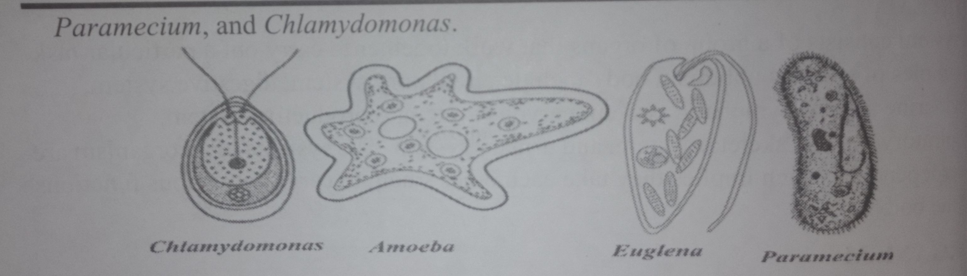
There is a progressive complexity in structure, function, activity, behavior, and mode of life from the simplest unicellular organism to the complex multicellular organism. Even though unicellular organisms can perform all life processes, they lack vital organs that are efficient to ensure their survival.
Living cells exist in two forms:
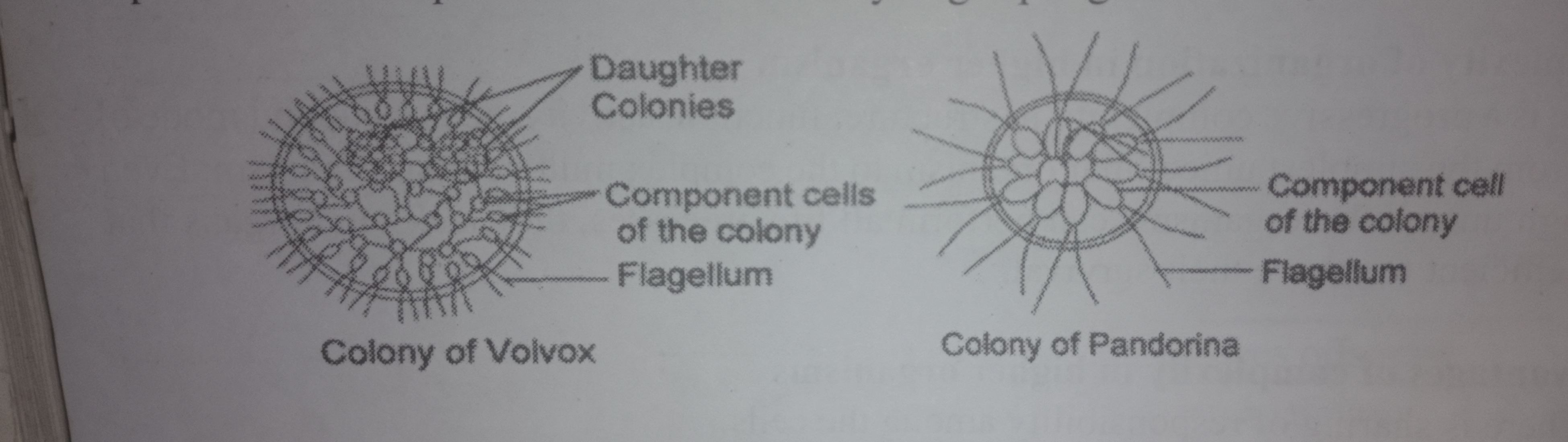
b. A colony is a loosely organized association of many similar cells held together by materials secreted by the cells themselves. This aggregation of independent cells or protists is called a colony. Examples include Sponges, Volvox, and Pandorina.
c. As filament- Certain cells are organized into filaments in which identical cells are joined end to end to form unbranched filaments. Each cell functions as an inde pendent living cell. Such organisms are multicellular and therefore exist as fila- ment. Examples are the Spirogyra, Zyguema, Oscillateria and Oedoqouium.

In multicellular organisms, a group of numerous, similar cells arranged together and performing a specific function is called a tissue. A group of similar tissues forming a layer in an organism which performs a specific function is called an organ. A group of organs which work together to perform specific functions are called a system. It is deduced that cells lead to tissues, tissues lead to organs, while organs lead to systems.
| SN. | Colonial Organism | Filamentous Organism |
|---|---|---|
| i | There is absence of intercellular wall | There is presence of intercellular wall |
| ii | Cells are connected by cytoplasmic material i.e. physiologically dependent | All cells are physiologically independent |
| iii | The identical cells form a mass | The identical cells form end-to-end arrangement in linear form |
| Examples | Volvox, Pandorina | Spirogyra, Zygnema, Oscillatoria |
Human body tissue consists of groups of cells with a similar structure working together for a specific function. There are four main types of tissue in a body:
Connective tissue connects or separates groups of other tissues. It is found in between all the other tissues and organs in the body. Connective tissue is made up of cells and ground substance, which is a gel that surrounds cells. Most connective tissue, except for lymph and blood, also contains fibers, which are long, narrow proteins. Fibers can be collagenous, which bind bones to tissues; elastic, which allow organs like the lungs to move; or reticular, which provide physical support to cells. Connective tissue also allows oxygen to diffuse from blood vessels into cells.
Muscle tissue comprises all the muscles in the body, and the specialized nature of the tissue is what allows muscles to contract. There are three types of muscle tissue: skeletal muscle, smooth muscle, and cardiac muscle.
Skeletal muscle anchors tendons to bones and allows the body to move. Cardiac muscle is found in the heart and contrach to pump blood. Smooth muscle is found in the intestines, where it helps move food through the digestive tract, and it is also found in other organs like blood vessels, the uterus, and the bladder. Skeletal and cardiac muscles are striated; this means that they contain sarcomeres (a unit of muscle tissue) that are arranged in a uniform pattern. Smooth muscle does not have sarcomeres.
Nervous tissue is found in the brain, spinal cord, and peripheral nerves, which are all parts of the nervous system. It is made up of neurons, which are nerve cells, and neuroglia, which are cells that help nerve impulses travel. Nervous tissue is grouped inte four types: gray matter and white matter in the brain, and nerves and ganglia in the peripheral nervous system. The main difference between gray and white matter is tha axons of the neurons in gray matter are unmyelinated, while white matter is myelinat ed.
Epithelial tissues are thin tissues that cover all the exposed surfaces of the body. They form the external skin, the inner lining of the mouth, digestive tract, secretory glands. the lining of hollow parts of every organ such as the heart, lungs, eyes, ears, the urogenital tract, as well as the ventricular system of the brain and central canals of the spinal cord.
It creates a barrier that helps protect organs, and it also has roles in absorbing water and nutrients, getting rid of waste, and secreting enzymes or hormones.
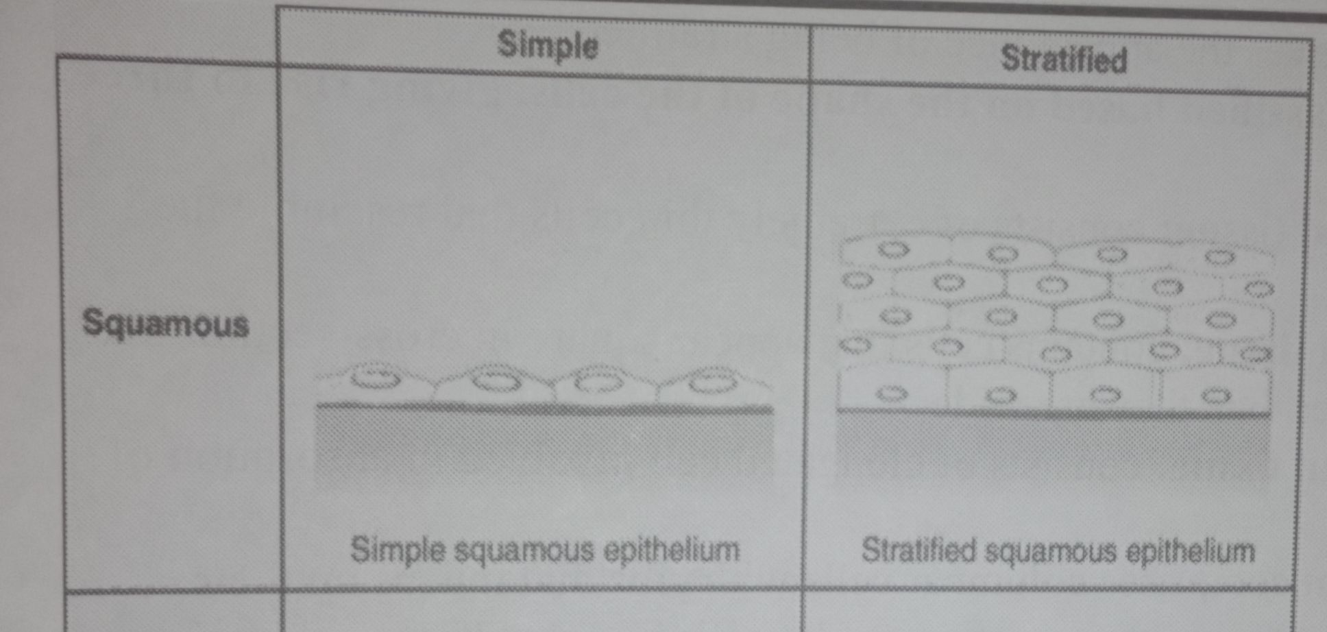
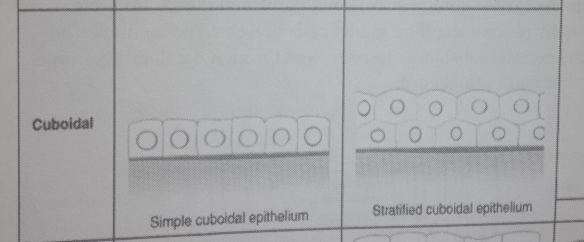
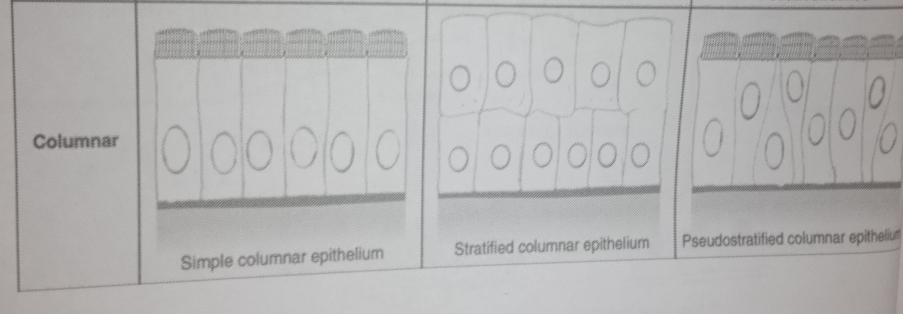
There are different types of epithelial tissue depending on their function in a particular location. The simplest classification of these tissues is based on the number of cell layers:
When the epithelium is composed of a single layer of cells, it is called simple epithelial tissue and those containing two or more layers of cells are called stratified epithelial tissues. One particular type is called pseudostratified because a single layer of cells having varying heights gives the appearance of being stratified.
Ciliated epithelium comprises columnar cells with cilia and mucus on their surfaces. There are many mucus-secreting goblet cells present. The combination of the cilia and mucus permits substances to be moved through the duct. Example is in the oviduct, trachea and spinal cord.
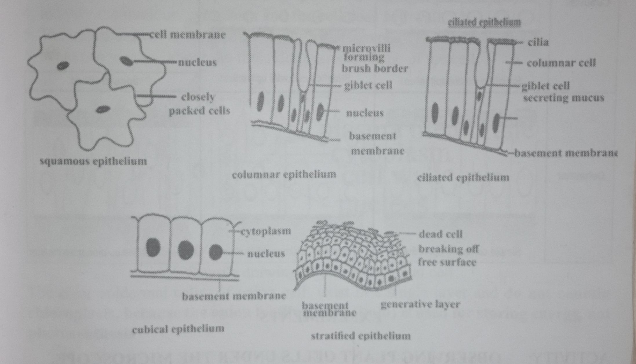
The number of cell layers and cell types together give rise to 6 different types of epithelial tissue.
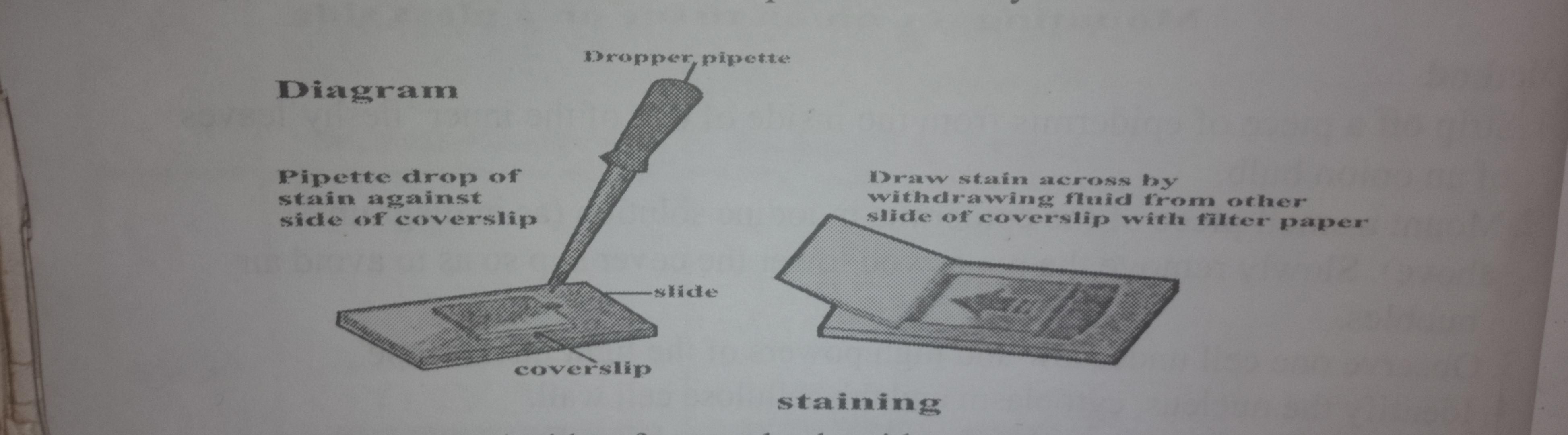
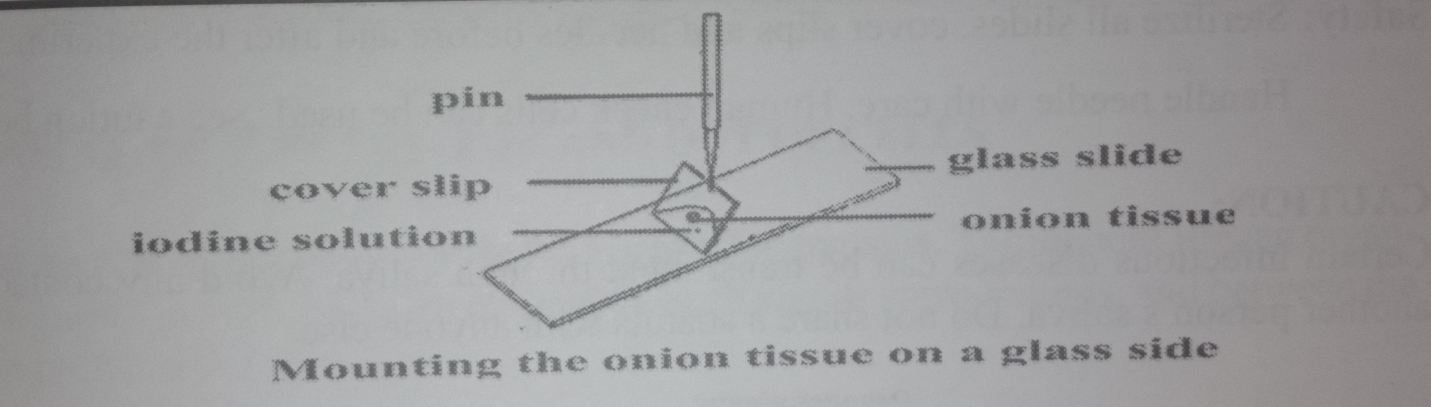
Safety: Handle needle & fragile coverslips with care
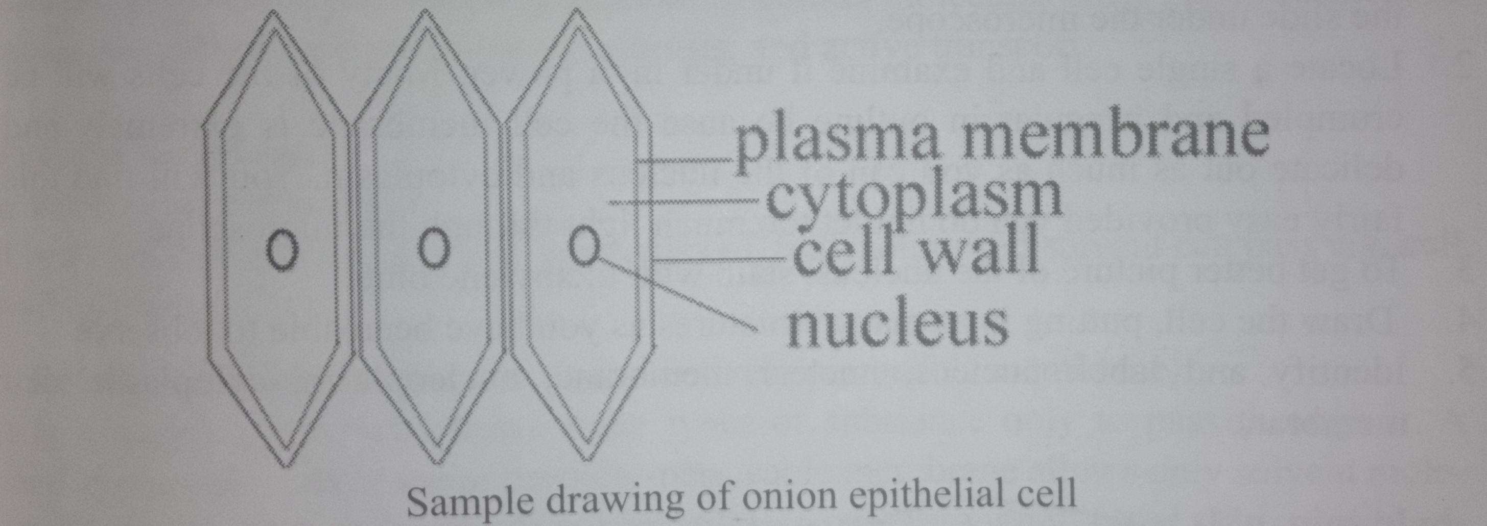
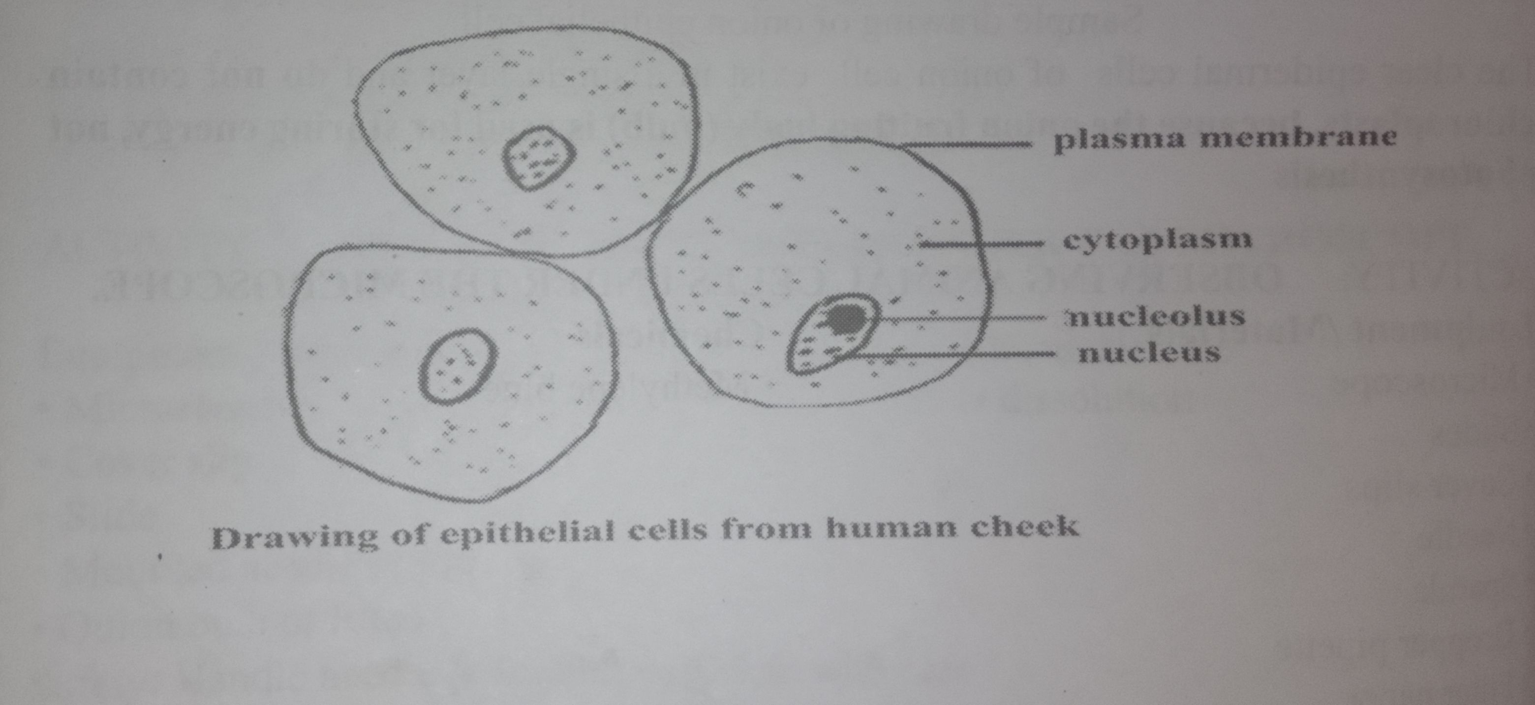
The clear epidermal cells of onion cell exist in a single layer and do not contain chloroplasts, because the onion fruiting body (bulb) is used for storing energy, not photosynthesis